Microscope are instruments designed to produce magnified visual or photographic
images of objects too small to be seen with the naked eye.
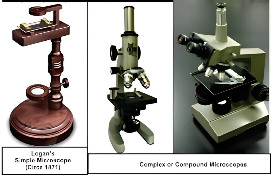
The first compound microscope was developed by Zacharias Janssenin Holland 1590.He designed a simple hollow tube with lenses at each end, and its magnification ranged 3X to 9X. Later on, Robert Hook improved his version for compound microscope.
The use of a microscope is called microscopy.
The two important parameters of microscopy are:
It is defined as the smallest distance between two points on a specimen that can still be distinguished as two objects.
There are two types of microscopes used in microscopy.
A microscope that uses visible light as a source of illumination and consists of lenses that magnify the light reflected from the object is called a light microscope.
A light microscope uses visible light as a source of illumination.


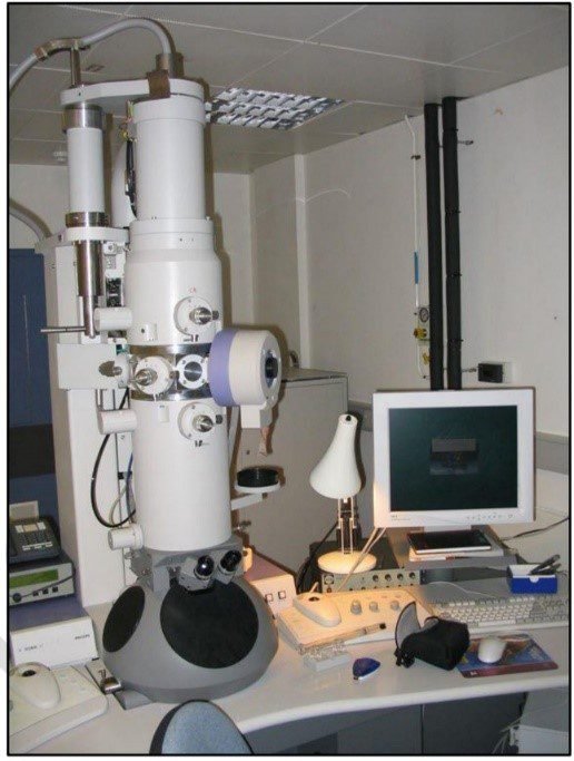
Microscopes that produce an image of a specimen by using a beam of electrons is called electron microscope.

Electron microscope has a resolution as small as 0.2 nanometer (nm).
Electron microscopes have a magnification up to 250,000 times.
A live cell cannot be imaged by an electron microscope.
There are two major types of electron microscopes:
Transmission electron microscopy (TEM) is used to obtain detailed images of internal structure of cells. The specimen is cut into extremely thin slices before imaging, an electron beam passes through the slice rather than skimming over its surface.
In scanning electron microscopy (SEM) a beam of electrons moves back and forth across the surface of a cell or tissue, creating a detailed image of the 3D surface.


0 of 10 Questions completed
Questions:
You have already completed the quiz before. Hence you can not start it again.
Quiz is loading…
You must sign in or sign up to start the quiz.
You must first complete the following:
0 of 10 Questions answered correctly
Your time:
Time has elapsed
You have reached 0 of 0 point(s), (0)
Earned Point(s): 0 of 0, (0)
0 Essay(s) Pending (Possible Point(s): 0)
What are microscopes designed to produce?
Who developed the first compound microscope?
What is the term for the use of a microscope?
What are the two important parameters of microscopy?
Which type of microscope uses visible light as a source of illumination?
What is the definition of a light microscope?
How does a light microscope produce a magnified image?
What is the resolution of a light microscope?
Which microscope is used to obtain detailed images of internal cell structures?
Which type of electron microscope moves back and forth across the surface of a cell, creating a detailed 3D image?